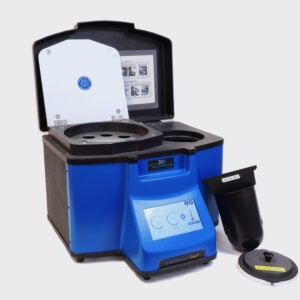
Tissue Homogenizer & Homogenization Protocol

Ideal for Tissue Samples

Save Time, Effort and Get Superior Results with

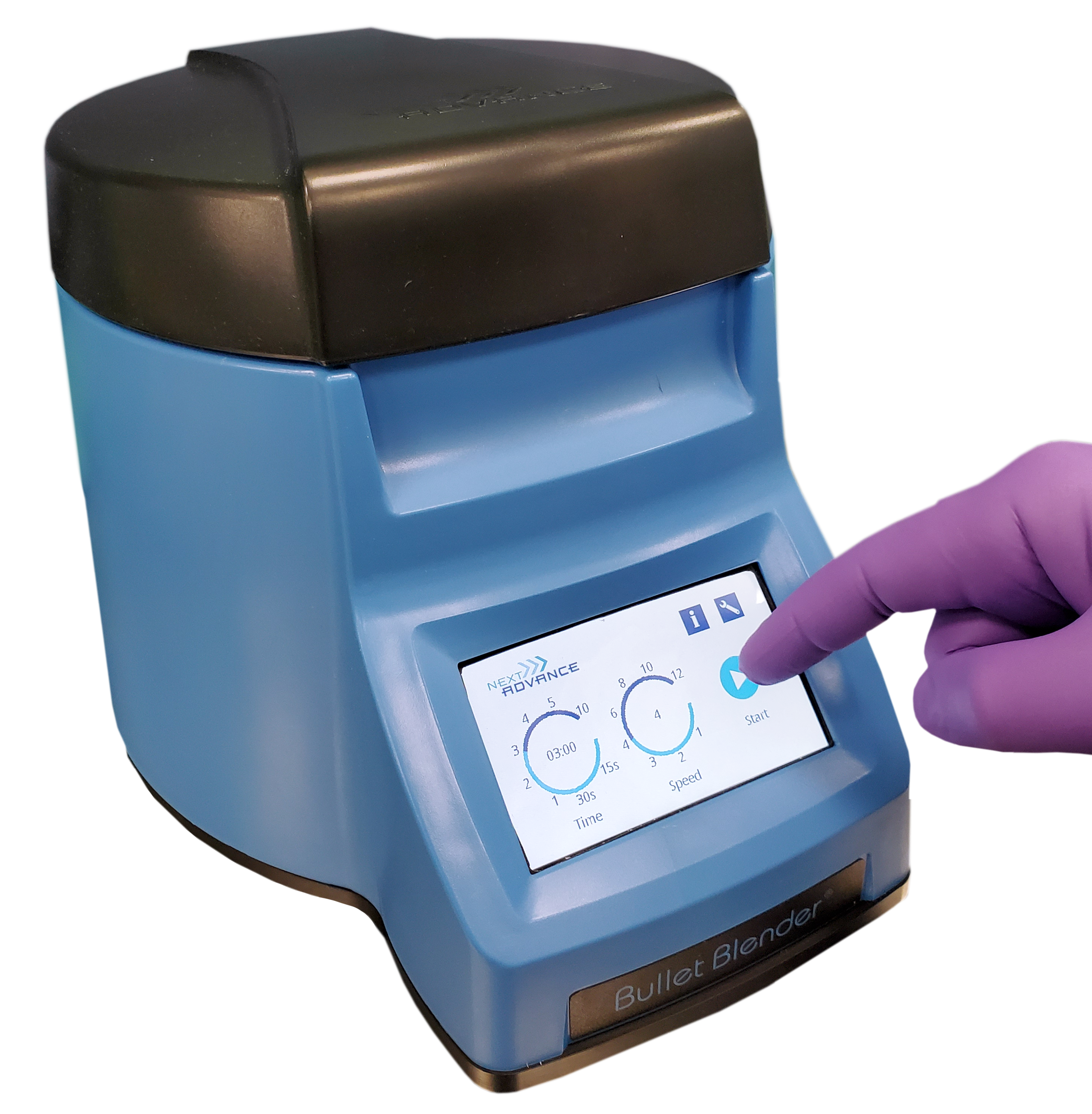
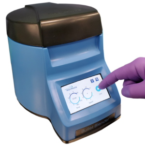
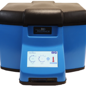
The Bullet Blender® Homogenizer

  • Consistent Results
  • Samples Stay Cool
  • No Cross Contamination
  • Easy & Convenient
  • Risk Free

Do you spend lots of time and effort homogenizing tissue samples? The Bullet Blender® is a multi-sample homogenizer that delivers high quality and superior yields. No other homogenizer comes close to delivering the Bullet Blender’s winning combination of top-quality performance and budget-friendly affordability.

Consistent and High Yield Results

Run up to 24 samples at the same time under microprocessor-controlled conditions, ensuring experimental reproducibility and high yield. Process samples from 10mg or less up to 3.5g.

No Cross Contamination

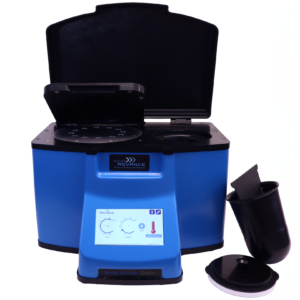
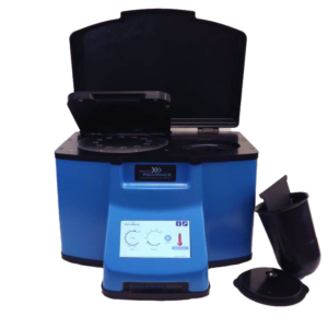
No part of the Bullet Blender® ever touches the tissue – the sample tubes are kept closed during homogenization. There are no probes to clean between samples.

Samples Stay Cool

Homogenizing causes only a few degrees of heating. Our Gold models keep samples at 4°C.

Easy and Convenient to Use

Just place beads and buffer along with your tissue sample in standard tubes, load tubes directly in the Bullet Blender, select time and speed, and press start.

Risk Free Purchase

The Bullet Blender® comes with a 30 day money back guarantee and a 2 year warranty, with a 3 year warranty on the motor. The simple, reliable design enables the Bullet Blenders to sell for a fraction of the price of ultrasonic or other agitation based instruments, yet provides an easier, quicker technique.

Example Bullet Blender settings for Tissue Homogenization

Sample size

See the Protocol

microcentrifuge tube model (up to 300 mg) Small adipose tissue samples
5mL tube model (100mg - 1g) Medium adipose tissue samples
50mL tube model (100mg - 3.5g) Large adipose tissue samples
the Bullet Blender high-throughput tissue homogenizer
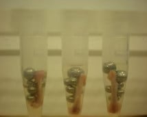
Three tubes with unhomogenized mouse femur.
Mouse Femur (Before)
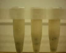
Three tubes with homogenized mouse femur.
Mouse Femur Homogenized in the Bullet Blender

What can you homogenize in a Bullet Blender?

Adipose, Adrenal Gland, Aorta, Bladder, Blood Vessel, Bone, Brain, Cartilage, Cecum, Colon, Cranium, Diaphragm, Duodenum, Ear Punch, Eye, Feces, Fish Organs, Gallbladder, Genital Warts, Heart, Hypothalamus, Intestinal Mucosa, Intestine, Jejunum, Kidney, Larynx, Liver, Lung, Lymph Node, Marrow, Meconium, Mouse Femur and Tibia, Muscle, Nasal/Olfactory Mucosa, Oropharynx, Pancreas, Pharynx, Pituitary Gland, Placenta, Polyp, Salivary Gland, Skin, Spleen, Stomach, Tail Snips, Teeth, Testes, Thalamus, Thymus, Thyroid, Tongue, Trachea, Tumor, Umbilical Cord, Uterus

Don’t see your tissue? Want more guidance? Contact our application support scientists:
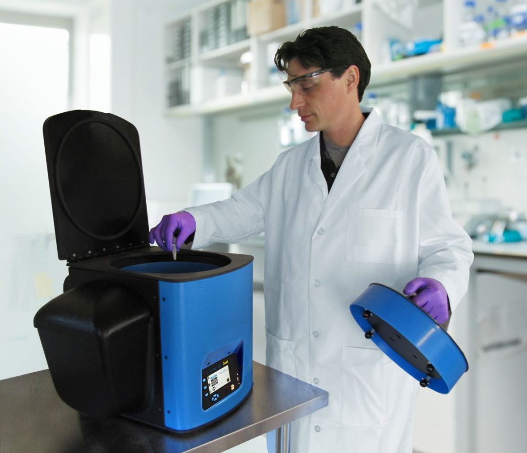


    Scientist using a Bullet Blender Gold

    Bullet Blender Models

    Selected publications for Tissue Homogenization​

    Clark, D. J., Mei, Y., Sun, S., Zhang, H., Yang, A. J., & Mao, L. (2016). Glycoproteomic Approach Identifies KRAS as a Positive Regulator of CREG1 in Non-small Cell Lung Cancer Cells. Theranostics, 6(1), 65–77. https://doi.org/10.7150/thno.12350
    Tranchemontagne, Z. R., Camire, R. B., O’Donnell, V. J., Baugh, J., & Burkholder, K. M. (2016). Staphylococcus aureus Strain USA300 Perturbs Acquisition of Lysosomal Enzymes and Requires Phagosomal Acidification for Survival inside Macrophages. Infection and Immunity, 84(1), 241–253. https://doi.org/10.1128/IAI.00704-15
    Park, S. J., Lee, H. W., Kim, H.-R., Kang, C., & Kim, H. M. (2016). A carboxylesterase-selective ratiometric fluorescent two-photon probe and its application to hepatocytes and liver tissues. Chem. Sci., 7(6), 3703–3709. https://doi.org/10.1039/C5SC05001D
    Lee, D., Ahn, C., An, B.-S., & Jeung, E.-B. (2015). Induction of the Estrogenic Marker Calbindn-D9k by Octamethylcyclotetrasiloxane. International Journal of Environmental Research and Public Health, 12(11), 14610–14625. https://doi.org/10.3390/ijerph121114610
    Upadhyay, A., Fontes, F. L., Gonzalez-Juarrero, M., McNeil, M. R., Crans, D. C., Jackson, M., & Crick, D. C. (2015). Partial Saturation of Menaquinone in Mycobacterium tuberculosis : Function and Essentiality of a Novel Reductase, MenJ. ACS Central Science, 1(6), 292–302. https://doi.org/10.1021/acscentsci.5b00212
    Meyer, R. E., Chuong, H. H., Hild, M., Hansen, C. L., Kinter, M., & Dawson, D. S. (2015). Ipl1/Aurora-B is necessary for kinetochore restructuring in meiosis I in Saccharomyces cerevisiae. Molecular Biology of the Cell, 26(17), 2986–3000. https://doi.org/10.1091/mbc.E15-01-0032
    Giengkam, S., Blakes, A., Utsahajit, P., Chaemchuen, S., Atwal, S., Blacksell, S. D., Paris, D. H., Day, N. P. J., & Salje, J. (2015). Improved Quantification, Propagation, Purification and Storage of the Obligate Intracellular Human Pathogen Orientia tsutsugamushi. PLOS Neglected Tropical Diseases, 9(8), e0004009. https://doi.org/10.1371/journal.pntd.0004009
    Dragunow, M., Feng, S., Rustenhoven, J., Curtis, M., & Faull, R. (2015). Studying Human Brain Inflammation in Leptomeningeal and Choroid Plexus Explant Cultures. Neurochemical Research. https://doi.org/10.1007/s11064-015-1682-2
    Ran, L., Yu, Q., Zhang, S., Xiong, F., Cheng, J., Yang, P., Xu, J.-F., Nie, H., Zhong, Q., Yang, X., Yang, F., Gong, Q., Kuczma, M., Kraj, P., Gu, W., Ren, B.-X., & Wang, C.-Y. (2015). Cx3cr1 deficiency in mice attenuates hepatic granuloma formation during acute schistosomiasis by enhancing the M2-type polarization of macrophages. Disease Models & Mechanisms, 8(7), 691–700. https://doi.org/10.1242/dmm.018242
    Sahu, R. (2015). Expression of the platelet-activating factor receptor enhances benzyl isothiocyanate-induced apoptosis in murine and human melanoma cells. Molecular Medicine Reports. https://doi.org/10.3892/mmr.2015.3371
    Behnia, F., Peltier, M. R., Saade, G. R., & Menon, R. (2015). Environmental Pollutant Polybrominated Diphenyl Ether, a Flame Retardant, Induces Primary Amnion Cell Senescence. American Journal of Reproductive Immunology, 74(5), 398–406. https://doi.org/10.1111/aji.12414
    Lydic, T. A., Townsend, S., Adda, C. G., Collins, C., Mathivanan, S., & Reid, G. E. (2015). Rapid and comprehensive ‘shotgun’ lipidome profiling of colorectal cancer cell derived exosomes. Methods, 87, 83–95. https://doi.org/10.1016/j.ymeth.2015.04.014
    Ozgul, S., Kasap, M., Akpinar, G., Kanli, A., Güzel, N., Karaosmanoglu, K., Baykal, A. T., & Iseri, P. (2015). Linking a compound-heterozygous Parkin mutant (Q311R and A371T) to Parkinson’s disease by using proteomic and molecular approaches. Neurochemistry International, 85–86, 1–13. https://doi.org/10.1016/j.neuint.2015.03.007
    Lemon, D. D., Harrison, B. C., Horn, T. R., Stratton, M. S., Ferguson, B. S., Wempe, M. F., & McKinsey, T. A. (2015). Promiscuous actions of small molecule inhibitors of the protein kinase D-class IIa HDAC axis in striated muscle. FEBS Letters, 589(10), 1080–1088. https://doi.org/10.1016/j.febslet.2015.03.017
    Mouton, J., Loos, B., Moolman-Smook, J. C., & Kinnear, C. J. (2015). Ascribing novel functions to the sarcomeric protein, myosin binding protein H (MyBPH) in cardiac sarcomere contraction. Experimental Cell Research, 331(2), 338–351. https://doi.org/10.1016/j.yexcr.2014.11.006
    Zhang, Z., He, L., Hu, S., Wang, Y., Lai, Q., Yang, P., Yu, Q., Zhang, S., Xiong, F., Simsekyilmaz, S., Ning, Q., Li, J., Zhang, D., Zhang, H., Xiang, X., Zhou, Z., Sun, H., & Wang, C.-Y. (2015). AAL exacerbates pro-inflammatory response in macrophages by regulating Mincle/Syk/Card9 signaling along with the Nlrp3 inflammasome assembly. American Journal of Translational Research, 7(10), 1812–1825.
    Shemesh, A., Wang, Y., Yang, Y., Yang, G.-S., Johnson, D. E., Backer, J. M., Pessin, J. E., & Zong, H. (2014). Suppression of mTORC1 activation in acid- -glucosidase-deficient cells and mice is ameliorated by leucine supplementation. AJP: Regulatory, Integrative and Comparative Physiology, 307(10), R1251–R1259. https://doi.org/10.1152/ajpregu.00212.2014
    Offer, S. M., Fossum, C. C., Wegner, N. J., Stuflesser, A. J., Butterfield, G. L., & Diasio, R. B. (2014). Comparative functional analysis of DPYD variants of potential clinical relevance to dihydropyrimidine dehydrogenase activity. Cancer Research, 74(9), 2545–2554. https://doi.org/10.1158/0008-5472.CAN-13-2482
    Da-Rè, C., Franzolin, E., Biscontin, A., Piazzesi, A., Pacchioni, B., Gagliani, M. C., Mazzotta, G., Tacchetti, C., Zordan, M. A., Zeviani, M., Bernardi, P., Bianchi, V., De Pittà, C., & Costa, R. (2014). Functional Characterization of d rim2 , the Drosophila melanogaster Homolog of the Yeast Mitochondrial Deoxynucleotide Transporter. Journal of Biological Chemistry, 289(11), 7448–7459. https://doi.org/10.1074/jbc.M113.543926
    Caruso, V., Hägglund, M. G., Badiali, L., Bagchi, S., Roshanbin, S., Ahmad, T., Schiöth, H. B., & Fredriksson, R. (2014). The G protein-coupled receptor GPR162 is widely distributed in the CNS and highly expressed in the hypothalamus and in hedonic feeding areas. Gene, 553(1), 1–6. https://doi.org/10.1016/j.gene.2014.09.042
    Henrich, M., Huber, K., Rydzewski, L., Kirsten, S., Spengler, B., Römpp, A., & Reinacher, M. (2014). Identification of T cell receptor signaling pathway proteins in a feline large granular lymphoma cell line by liquid chromatography tandem mass spectrometry. Veterinary Immunology and Immunopathology, 161(1–2), 116–121. https://doi.org/10.1016/j.vetimm.2014.06.004
    Bartnikowski, M., Klein, T. J., Melchels, F. P. W., & Woodruff, M. A. (2014). Effects of scaffold architecture on mechanical characteristics and osteoblast response to static and perfusion bioreactor cultures: Scaffold Architecture Static Perfusion Bioreactor. Biotechnology and Bioengineering, 111(7), 1440–1451. https://doi.org/10.1002/bit.25200
    Menon, R., Boldogh, I., Urrabaz-Garza, R., Polettini, J., Syed, T. A., Saade, G. R., Papaconstantinou, J., & Taylor, R. N. (2013). Senescence of Primary Amniotic Cells via Oxidative DNA Damage. PLoS ONE, 8(12), e83416. https://doi.org/10.1371/journal.pone.0083416
    Townsend, K. L., An, D., Lynes, M. D., Huang, T. L., Zhang, H., Goodyear, L. J., & Tseng, Y.-H. (2013). Increased Mitochondrial Activity in BMP7-Treated Brown Adipocytes, Due to Increased CPT1- and CD36-Mediated Fatty Acid Uptake. Antioxidants & Redox Signaling, 19(3), 243–257. https://doi.org/10.1089/ars.2012.4536
    Thoene, J., Goss, T., Witcher, M., Mullet, J., N’Kuli, F., Van Der Smissen, P., Courtoy, P., & Hahn, S. H. (2013). In vitro correction of disorders of lysosomal transport by microvesicles derived from baculovirus-infected Spodoptera cells. Molecular Genetics and Metabolism, 109(1), 77–85. https://doi.org/10.1016/j.ymgme.2013.01.014
    Thomas, S. N., Waters, K. M., Morgan, W. F., Yang, A. J., & Baulch, J. E. (2012). Quantitative proteomic analysis of mitochondrial proteins reveals prosurvival mechanisms in the perpetuation of radiation-induced genomic instability. Free Radical Biology and Medicine, 53(3), 618–628. https://doi.org/10.1016/j.freeradbiomed.2012.03.025
    Willis, M. N., Liu, Y., Biterova, E. I., Simpson, M. A., Kim, H., Lee, J., & Barycki, J. J. (2011). Enzymatic Defects Underlying Hereditary Glutamate Cysteine Ligase Deficiency Are Mitigated by Association of the Catalytic and Regulatory Subunits. Biochemistry, 50(29), 6508–6517. https://doi.org/10.1021/bi200708w
    Wu, S., Wang, L., Guo, W., Liu, X., Liu, J., Wei, X., & Fang, B. (2011). Analogues and Derivatives of Oncrasin-1, a Novel Inhibitor of the C-Terminal Domain of RNA Polymerase II and Their Antitumor Activities. Journal of Medicinal Chemistry, 54(8), 2668–2679. https://doi.org/10.1021/jm101417n

    Have any questions? Ask us!​

    Success!

    The discount has been applied. You will see it when you checkout.

    There has been a problem

    Unfortunately this discount cannot be applied to your cart.